Pleural fluid is found in the pleural cavity and serves as a lubricant for the movement of the lungs during inhalation and exhalation. It is derived from a plasma filtrate from blood capillaries and is found in small quantities between the layers of the pleurae - membranes that cover the chest cavity and the outside of each lung.
A variety of conditions and diseases can cause inflammation of the pleurae (pleuritis) and/or excessive accumulation of pleural fluid (pleural effusion). Pleural fluid analysis comprises a group of tests used to determine the cause. There are two main reasons fluid may collect in the pleural space:
Differentiation between the types of fluid is important because it helps diagnose the specific disease or condition. Doctors and laboratory scientists use an initial set of tests (cell count, albumin and appearance of the fluid) to distinguish between transudates and exudates. Once the fluid is determined to be one or the other, additional tests may be performed to further pinpoint the disease or condition causing pleuritis and/or pleural effusion.
Pleural fluid analysis is used to help diagnose the cause of inflammation of the pleurae (pleuritis) and/or accumulation of fluid in the pleural space (pleural effusion). There are two main reasons for fluid accumulation, and an initial set of tests (albumin, cell count and appearance of the fluid) is used to differentiate between the two types of fluid that may be produced:
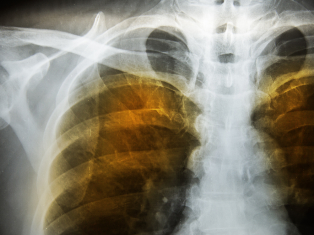
Pleural fluid analysis is requested after the information from a detailed history and physical examination, review of blood tests, chest imaging by X-ray and/or ultrasonography have been evaluated by the doctor.
An initial set of tests performed on a sample of pleural fluid helps determine whether the fluid is a transudate or exudate:
Transudate
Exudate
Physical characteristics
The normal appearance of a sample of pleural fluid is usually light yellow and clear. Abnormal results may give clues to the conditions or diseases present and may include:
Chemical tests
Tests that may be performed in addition to protein or albumin may include:
Microscopic examination
Normal pleural fluid has small numbers of white blood cells (WBCs) but no red blood cells (RBCs) or microorganisms. Laboratories may examine the pleural fluid and/or use a special centrifuge (cytocentrifuge) to concentrate the fluid’s cells on a slide. The slide is treated with a special stain and evaluated for the different kinds of cells that may be present.
Infectious disease tests
These tests may be performed to look for microorganisms if infection is suspected:
A blood glucose, protein, albumin or LD may be ordered to compare concentrations with those in the pleural fluid.
Thoracentesis is the removal of pleural fluid from the pleural cavity with a needle and syringe. The person is positioned sitting upright with arms raised and supported. A local anaesthetic is applied and then the doctor inserts the needle into the pleural cavity and the sample is removed.
Yes. Sometimes it will be performed to drain excess pleural fluid – to relieve pressure on the lungs. A catheter tube may be used to drain larger amounts of fluid and to drain recurrent fluid accumulations.